


Arthritis of the Hip (ATS) is a slowly destructive disease. Under the influence of a variety of reasons, the structure and properties of hyaline cartilage undergo irreversible changes during the development of the disease, resulting in increased pressure on the articular surface, deformation or fusion. The joint of the hip is often affected by arthrosis, given that mechanical overload is considered one of the main causes of disease development.
Anatomical features of the hip joint
The hip joint (TC) is the junction of the pelvis and femur. This joint reduces and expands the lower extremities, lifts the legs and draws them over the body, and performs walking motions. From birth to life, a person's hip joints are under a lot of load.
From the side of the pelvic bone, the "acetabular" cavity participates in the joint, and from the side of the femur, its epiphysis. The edge of the acetabulum has a collagen lip that acts as a sort of gasket, holding the epiphysis of the femur tightly in its depression. The hollow in the center of the acetabulum is covered with a collagen membrane and is where the femoral ligament attaches.
The composition of TS capsules includes ligaments:
- femur-ilium - the strongest ligament, which can withstand loads of more than 200 kg and prevents excessive rearward arching of the hips;
- femur - pubic bone - responsible for the abduction and reduction of the thigh, thereby limiting its circular motion;
- femoral ischia - protects the vehicle from concussions, reduces loads during walking and running;
- Circle (Ring) - Prevents dislocation and secures the femoral head in the pelvic cavity, is the basis of the joint capsule.
A number of muscle groups and tendons allow the vehicle to move about three axes:
- portrait (vertical).
- Landscape (horizontal, frontal).
- Sagittal plane (anterior-posterior).
Arthropathy can occur in healthy joints or as a continuation of an existing disorder of the musculoskeletal system.
What kind of disease is this?
Hyaline cartilage acts as a shock absorber and protects joint surfaces from damage. ATS is a progressive disease in which the structure of collagen cartilage fibers changes, leading to their subsequent fragmentation and destruction. Fragments of cartilage fibers can cause an inflammatory process if they get into the joint cavity. Exposed surfaces can cause changes in bone tissue due to increased friction and pressure. Compensatory growth of residual cartilaginous tissue along the epiphyseal margin with subsequent ossification results in ankylosis (immobilization of bony junctions). In later stages, without proper treatment, the patient becomes completely incapacitated and becomes disabled. Destructive processes are caused by various reasons.
There are the following types of arthropathy in the hip joint:
- basic. Its cause is not fully understood. Idiopathic (primary) arthropathy develops in previously healthy joints. Most often, it occurs in older people.
- middle school. It is caused by previous joint organ diseases, congenital abnormalities, changes in the work of organs and human vital activity systems.
The disease develops in one joint or affects both joints at the same time.
cause of disease
Among the causes of the appearance of the disease and its progression, the following factors were identified:
- Genetic susceptibility to disease development.
- Bone and joint injuries (dislocations, fractures, sprains and tendons).
- Unbearable systemic strength and physical activity.
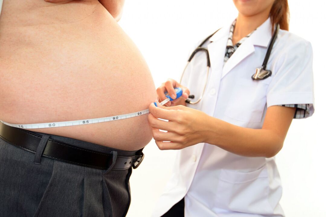
- overweight.
- Endocrine system dysfunction (diabetes, psoriasis).
- Congenital pathologies of musculoskeletal structure and development.
- Professional features of labor activity.
- Poor local circulation.
- Previous diseases caused by pathogenic bacteria.
- Legg-Calve-Perthes disease.
- Metabolic disorders (gout).
- lack of exercise.
- immune disease.
These causes do not always cause ATS. Most often, the activation of pathological processes can be caused by:
- Increased stress and physical activity;
- often overworked;
- vehicle or whole body hypothermia;
- suddenly lift a heavy object;
- hormone imbalance;
- radiation exposure.
disease symptoms
The symptoms of ATS are similar to those of arthropathy in other joints.
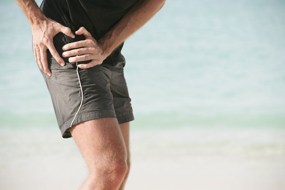
The main characteristic symptoms of the disease are considered to be:
- Stiffness in the morning or after prolonged immobility.
- Range of motion is reduced and gait changes.
- Pain, first caused by mechanical or physical pressure, then persists.
- Creaking, creaking, and clicking manifestations of sudden movements.
- The affected limb is visibly lame.
- The development of contractures (limitation of passive movement).
- Narrowing or closing of the joint space (x-ray signs).
The severity of hip arthritis symptoms depends on how advanced the disease is and how well the patient's body responds.
Stages of Hip Disease
On the basis of clinical presentation, hip arthropathy can be divided into 4 stages:
- Grade 1 arthropathy of the hip joint without obvious pain and other manifestations. This stage is difficult to diagnose, and the disease can be detected using biochemical studies of the hyaline cartilage tissue and determination of insufficient amounts of glycosaminoglycans. Patients experience joint pain when they begin physical activity and rarely feel pain.
- Second-degree arthropathy of the hip is characterized by changes in the density and elasticity of the cartilage fibers. Cracks and breaks appear. The depreciation function decreases. The pain intensified, radiated to the groin area, the affected limb was diluted, and the reset movement was limited.
- In the third degree, delamination of the cartilage fibers occurs with greater intensity. The articular surface bears excessive pressure, forming local ischemic foci. Cartilaginous tissue grows along the edges of the epiphysis. Pain at damaged bony junctions is independent of activity and rest. Joints "creak" and "sit-ups" when performing any movement. The range of motion is reduced in all axes.
- Fourth degree is characterized by exposure of the surface of joint components and formation of ulcers and depressions. Poor fixation of the femoral head in the acetabulum, resulting in violation of the relative separation of the articular surfaces. During this time, patients experience excruciating pain due to narrowing (and sometimes closing) of the joint space and compression of nerve fiber bundles and blood vessels. Movement is restricted, sometimes completely.
Classification of pathological changes caused by ATS is necessary to understand the mechanisms and characteristics of disease development. Determining the severity of the disease helps determine the correct treatment strategy and disability (in the case of severe disease).
possible consequences
The progression of ATS leads not only to deformation of the femoral head and pelvic cavity, but also to the development of a pathological process of the function of the entire joint apparatus.
Pathology caused by complications of hip arthrosis:
- Synovitis (inflammation of the synovial membrane of a joint);
- Aseptic necrosis of the femoral head;
- joint destruction (osteonecrosis);
- Inflammation of the joint capsule and changes in the amount of synovial fluid;
- Partial or complete ankylosis (fixation of bones and joints);
- Contractures (limited movement and inability to flex or extend).
The development of ATS complications invariably leads to a deterioration in the patient's general condition, quality of life, and loss of exercise capacity without assistance.
diagnosis method
Diagnosis of hip arthropathy in the initial stages is difficult. Symptoms become apparent only when the epiphysis and nerve fibers of the bone are involved in the pathological process.
During the physical examination in the progressive phase, the following are noted:
- visual changes in joint contours;
- soreness on palpation;
- Sometimes swelling of the tissue around the joint;
- The affected limb is shortened.
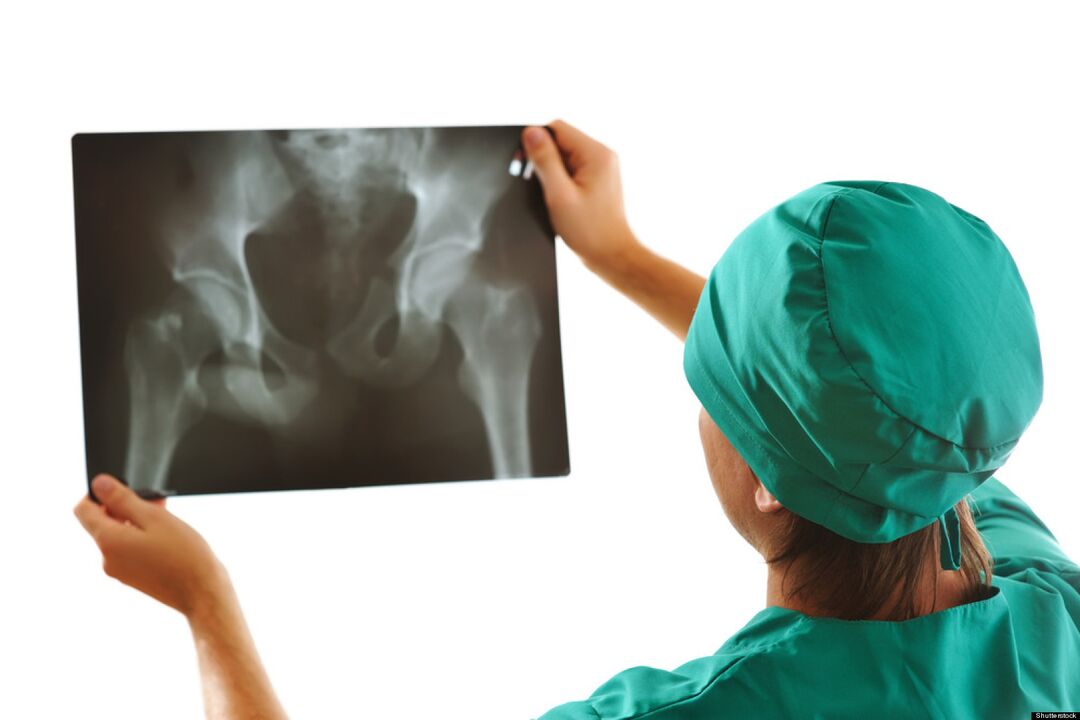
The main function of ATS diagnosis is X-ray examination. Use as an auxiliary diagnostic method:
- Ultrasound, magnetic resonance imaging.
- CT scan.
- Joint lubrication puncture (synovial fluid).
- Diagnosis is made using an arthroscope (microprobe).
- Clinical and biochemical laboratory tests of urine and blood.
Timely diagnosis improves treatment outcomes and prolongs patient life.
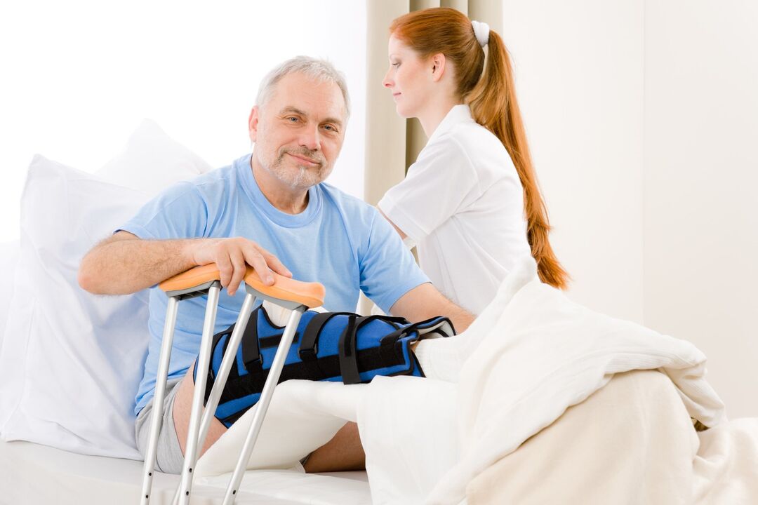
How do I apply for a disability?
It is impossible to completely cure the disease. In order to confirm the entitlement to social benefits and to assign a disability group after an examination by a narrow specialist, you must contact your doctor.

Indications for assigning disability in the case of hip arthrosis are:
- Oligoarthropathy (damage to no more than 2 joints) TS 2 degrees;
- Combined TS 2nd degree arthropathy and knee joint 3rd degree arthropathy;
- A reduction in the length of the affected limb of more than 6 cm;
- React Fluid's automatic telephone exchange, for the record.
Identifying disability groups will help:
- Carefully collect medical records;
- the conclusions of the Medical Advisory Committee (MCC);
- the results of diagnostic studies;
- Through the Medical and Social Experts Committee (MSEC).
If the expert committee's decision is negative, an appeal can be made to the higher authorities.
prevention
Preventive measures are an easy way to avoid the development of this disease. Preventive measures include:
- Stick to an active lifestyle.
- Control your body weight.
- Optimize nutrition and work and rest patterns.
- Reduce mechanical and physical load.
- Treatment of diseases of viral and infectious etiology.
- Prevention and prevention of injuries at home and work.
- Regular preventive inspections.
in conclusion
For the answer to the common question "Can hip joint disease be cured? ", experts gave a negative answer. Destroyed cartilage tissue cannot be fully restored, just as it is impossible to completely correct the deformation and destruction of bone in a joint. Not ignoring even minor manifestations of hip disease can reduce the chances of preventing further progression of the disease.